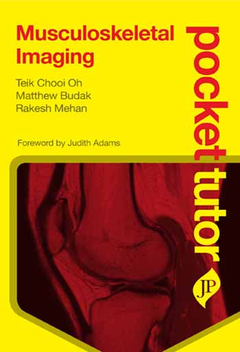
The book opens by demonstrating the appearance of normal tissues before going on to illustrate the radiological features of pathological tissues. Having provided a framework for recognising normal findings and key abnormal signs, subsequent chapters summarise the radiological anatomy, clinical appearance and management of the most common musculoskeletal diseases, by body region. A final chapter demonstrates common systemic pathologies which are not easily grouped into a single region. All chapters are lavishly illustrated with high-quality, clearly labelled images.
... Titles in the Pocket Tutor series give practical guidance on subjects that medical students and foundation doctors need help with ÔøΩon the goÔøΩ, at a highly affordable price that puts them within reach of those rotating through modular courses or working on attachment.
Highly structured, bite-size content helps novices combat the ÔøΩfear factorÔøΩ associated with day to day clinical training, and provides ÔøΩjust enoughÔøΩ for a new rotation.
ÔøΩ Highly affordable price and convenient pocket size format fits in back pocket!
ÔøΩ Logical, sequential content: the first principles of MSK imaging, then a guide to understanding a normal image and the building blocks of an abnormal image, before describing specific clinical disorders
ÔøΩ Clinical disorders are illustrated by high quality radiographs, ultrasounds, CTs and MRIs, with brief accompanying text that clearly identifies the defining features of the image
ÔøΩ Focuses on the conditions that medical students, junior doctors and trainees are most likely to see and be tested on